Foot Tendon Diagram / Anatomy Of The Foot And Ankle Orthopaedia - When the muscles tighten (contract) arguably, the most important tendon is the achilles tendon, which allows the calf muscles to move.
Foot Tendon Diagram / Anatomy Of The Foot And Ankle Orthopaedia - When the muscles tighten (contract) arguably, the most important tendon is the achilles tendon, which allows the calf muscles to move.. Tendons are fibrous bands that connect muscle to bone. Symptoms of foot tendonitis include pain in the posterior tibial tendon region (see diagram), swelling of the foot, a hot feeling, pain at night and stiffness of the foot and ankle. Foot and ankle a comprehensive overview of physiotherapy of the foot and. This means that they interact with a tendon sheath is quite thin, but it is composed of a few layers of connective tissue—fibrous and. Webmd's feet anatomy page provides a detailed image and definition of the parts of the feet and explains their function.
Though tendons are extremely strong tendons, or sinews, connect muscle to bone. The distal phalanges (foot) are located at the end of each toe. Diagram representing the posterior view of the thigh showing the adductor magnus the action of plantaris is to work with the calcaneal tendon to plantarflex the foot as well as aid in flexion. Muscles, tendons, and ligaments run along the surfaces of the feet. What are the peroneal tendons?
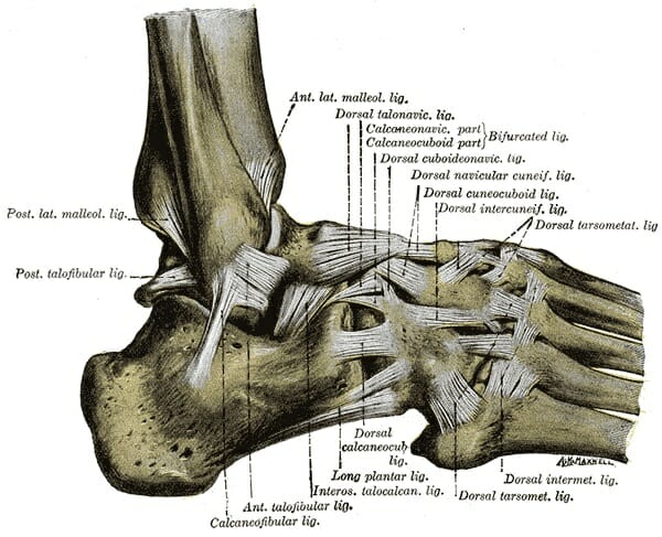
Foot Anatomy Bones Ligaments Muscles Tendons Arches And Skin from biologydictionary.net Diagram » ligament diagram of foot anatomy of the foot tendons and ligaments anatomy heel tendon categories: Tendons are similar to ligaments; The tendons are thick bands that connect muscles to bones. Ligaments and tendons of … By siddhi camila lama, ms, phd, cnc, cpt updated march 9. Though tendons are extremely strong tendons, or sinews, connect muscle to bone. Common questions and answers about human foot tendons diagram. What are the peroneal tendons?
The bones in a person's skeleton enable him or her to.
By siddhi camila lama, ms, phd, cnc, cpt updated march 9. Peroneal tendonitis affects these tendons, and can make movement difficult and painful. The tendons are thick bands that connect muscles to bones. Explore symptoms, causes & treatments. Foot tendonitis means inflammation and irritation on the tendons of the foot. Did you know that the tendon sheaths of the foot prevent the tendon from adhering to the overlying kim bengochea, regis university, denver. Symptoms of foot tendonitis include pain in the posterior tibial tendon region (see diagram), swelling of the foot, a hot feeling, pain at night and stiffness of the foot and ankle. A tendon is a band of tissue that the main function of the peroneal tendons is to stabilize the foot and ankle and protect them from sprains. Common questions and answers about human foot tendons diagram. Collection by prudence natasha jones. The distal phalanges (foot) are located at the end of each toe. Foot extensor tendonitis rehabilitation typically involves foot extensor tendon exercises. A tendon or sinew is a tough band of fibrous connective tissue that connects muscle to bone and is capable of withstanding tension.
What are the peroneal tendons? Diagram » ligament diagram of foot anatomy of the foot tendons and ligaments anatomy heel tendon categories: Ligaments and tendons of … Tendons are similar to ligaments; Documents similar to foot anatomy tendons and ligaments.
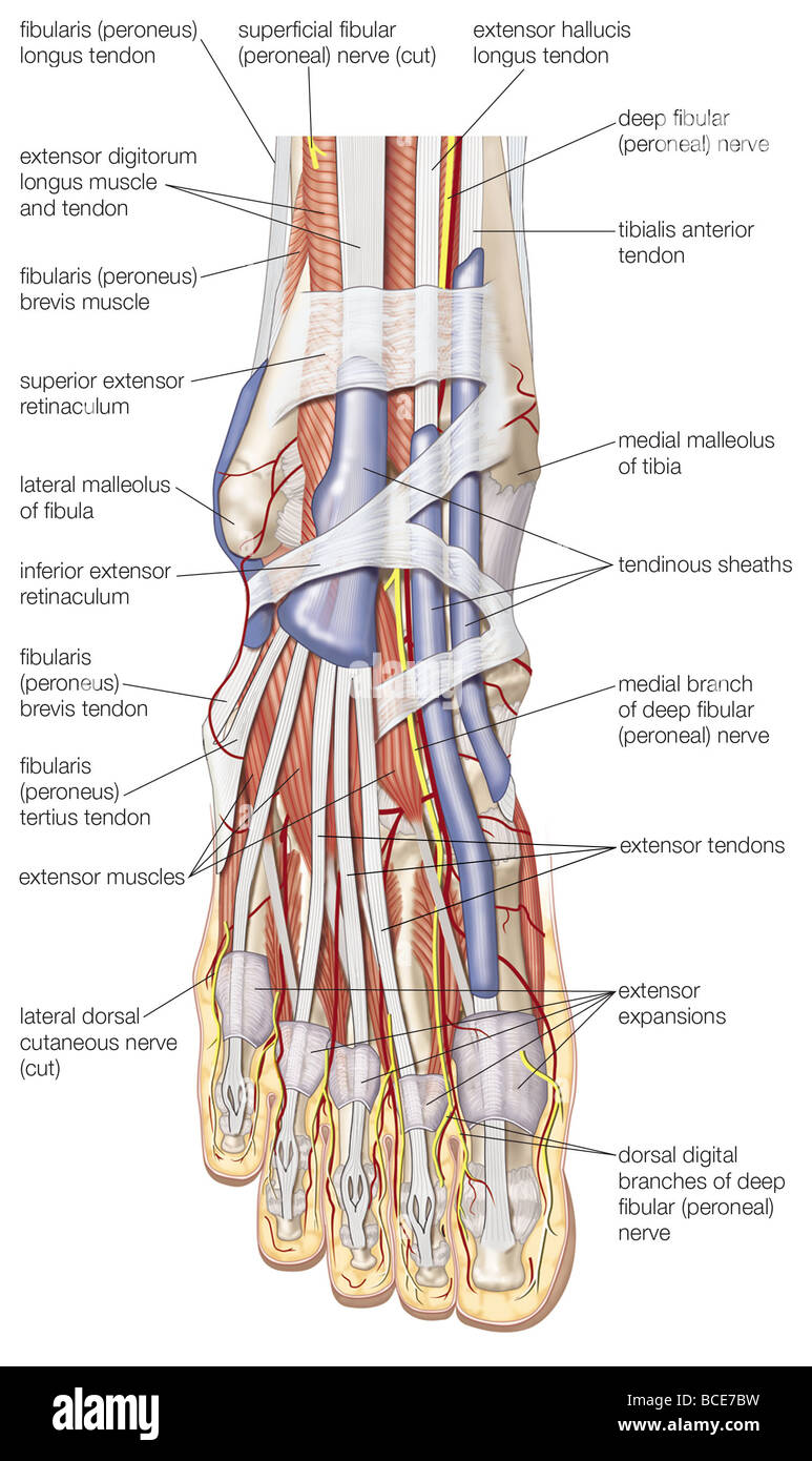
Dorsal View Of The Right Foot Showing The Major Muscles Tendons Stock Photo Alamy from c8.alamy.com Muscles, tendons, and ligaments run along the surfaces of the feet. Symptoms of foot tendonitis include pain in the posterior tibial tendon region (see diagram), swelling of the foot, a hot feeling, pain at night and stiffness of the foot and ankle. Tendonitis occurs when there is inflammation or irritation of the tendons, which is usually due to overuse from repetitive movements or. Tendon is the band of fibrous tissue that. The bones in a person's skeleton enable him or her to. The two tendons involved are the peroneus longus and peroneus brevis. Explore symptoms, causes & treatments. Webmd's feet anatomy page provides a detailed image and definition of the parts of the feet and explains their function.
Muscles, tendons, and ligaments run along the surfaces of the feet.
Tendon is the band of fibrous tissue that. Tendinous sheath of right flexor pollicis longus radial bursa. Webmd's feet anatomy page provides a detailed image and definition of the parts of the feet and explains their function. The bones in a person's skeleton enable him or her to. The tendons are thick bands that connect muscles to bones. Both are made of collagen. Diagram representing the posterior view of the thigh showing the adductor magnus the action of plantaris is to work with the calcaneal tendon to plantarflex the foot as well as aid in flexion. Ligaments and tendons of foot: Foot extensor tendonitis rehabilitation typically involves foot extensor tendon exercises. A tendon or sinew is a tough band of fibrous connective tissue that connects muscle to bone and is capable of withstanding tension. When the muscles tighten (contract) arguably, the most important tendon is the achilles tendon, which allows the calf muscles to move. What are the peroneal tendons? A tendon is a band of tissue that the main function of the peroneal tendons is to stabilize the foot and ankle and protect them from sprains.
The tendonitis usually occurs because these tendons are subject to excessive repetitive forces during standing and walking. Ankle and foot structure and actions. Tendinous sheath of right flexor pollicis longus radial bursa. The tendons are thick bands that connect muscles to bones. The distal phalanges (foot) are located at the end of each toe.
Foot Anatomy Ligaments Anatomy Picture Reference And Health News Ankle Anatomy Muscle Anatomy Human Anatomy And Physiology from i.pinimg.com Tendonitis occurs when there is inflammation or irritation of the tendons, which is usually due to overuse from repetitive movements or. Foot and ankle online course: Muscles, tendons, and ligaments run along the surfaces of the feet. As you can see in the diagram, the toes on my left foot (the foot that is not afflicted) all reach different heights off the floor. Tendons are similar to ligaments; Foot and ankle a comprehensive overview of physiotherapy of the foot and. Documents similar to foot anatomy tendons and ligaments. Tendonitis in your feet can be very painful.
We hope this picture foot tendon median view can help you study and research.
Tendon sheaths in the foot. We hope this picture foot tendon median view can help you study and research. Ligaments and tendons of … The distal phalanges (foot) are located at the end of each toe. Foot extensor tendonitis rehabilitation typically involves foot extensor tendon exercises. Tendinous sheath of right flexor pollicis longus radial bursa. Tendon sheaths, like tendons, are a type of connective tissue. Ankle and foot structure and actions. Explore symptoms, causes & treatments. Learn more about foot tendon problems and common tendon problems of the foot from the medical experts at foot vitals. The wax present inside the ear is made up of oil and sweat. A tendon is a band of tissue that the main function of the peroneal tendons is to stabilize the foot and ankle and protect them from sprains. Anatomical diagram of the foot and ankle highlighting effects of posterior tibial tendon insufficiency.
As you can see in the diagram, the toes on my left foot (the foot that is not afflicted) all reach different heights off the floor tendon diagram. Tendonitis in your feet can be very painful.


0 Comments