Labeled Diagram Of Shoulder Muscles : Shoulder Mri Radiographical And Illustrated Anatomical Atlas / Labeled anatomy chart of neck and shoulder muscles on, 3d rendering white background.
Labeled Diagram Of Shoulder Muscles : Shoulder Mri Radiographical And Illustrated Anatomical Atlas / Labeled anatomy chart of neck and shoulder muscles on, 3d rendering white background.. Tutorials on the shoulder muscles (e.g rotator cuff muscles: Shoulder flexion is movement of the shoulder in a forward motion. The muscles of the shoulder are associated with movements at the shoulder joint. Click on image for larger labeled, picture. An mri of the shoulder of a healthy subject was performed in the 3 planes of space (coronal, axial, sagittal) commonly used in osteoarticular imaging, with two weightings to explore the musculoskeletal.
In this video we'll explore the muscles and functions of the shoulder girdle (pectoral girdle). Shoulder muscles diagram includes some organs and can give you some detailed information as well as can be seen through shoulder anatomy diagram. They produce the characteristic shape of the shoulder, and can be divided into two groups Due to its location, it belongs to the chest. Test your knowledge in our quiz about the shoulder muscles.
The Back Muscle Anatomy Human Anatomy from graphdiagram.com This article describes the anatomy of the upper limb muscles including their attachments actions and location. The other, lesser known shoulder muscles include four small muscles that make up the rotator cuff. Supraspinatus, infraspinatus, ters minor,.et), using interactive animations and labeled diagrams. Why do shoulder problems affect the rest of the body? Many of the muscles that attach onto the shoulder girdle also attach onto the: The latissimus dorsi also transversely extends and flexes the. 9 in rehabilitation from such injuries context: The shoulder muscles include skeletal muscles that are attached to the head of the humerus which performs various direct and indirect functions of at the fixed end of the humerus, the pectoralis major muscle pulls the shoulder girdle in the ventral direction.
Click on image for larger labeled, picture.
The diagram above shows part a myofibril called a sarcomere. Specifically, the four rotator cuff muscles. The muscles of the shoulder are associated with movements at the shoulder joint. Muscles connecting upper limb to vertebral column muscles of shoulder. Shoulder flexion is movement of the shoulder in a forward motion. An example of shoulder flexion can be seen when reaching forward to grasp an object. This article describes the anatomy of the upper limb muscles including their attachments actions and location. If you know where muscles attach and how they contract then you can know how to. 6 photos of the shoulder muscles labelled diagram. The shoulder is not a single joint, but a complex arrangement of bones, ligaments, muscles, and tendons that is better bones of the shoulder girdle. 4 shoulder muscle imbalance may disturb the proximal stability and increase the incidence of distal joint injuries. Here we explain the major muscles of the human body. The malfunction of the proximal.
Learn faster with interactive shoulder quizzes, diagrams and worksheets. 6 photos of the shoulder muscles labelled diagram. Tutorials on the shoulder muscles (e.g rotator cuff muscles: Muscles connecting upper limb to vertebral column muscles of shoulder. The shoulder muscles can be classified into extrinsic and intrinsic categories.
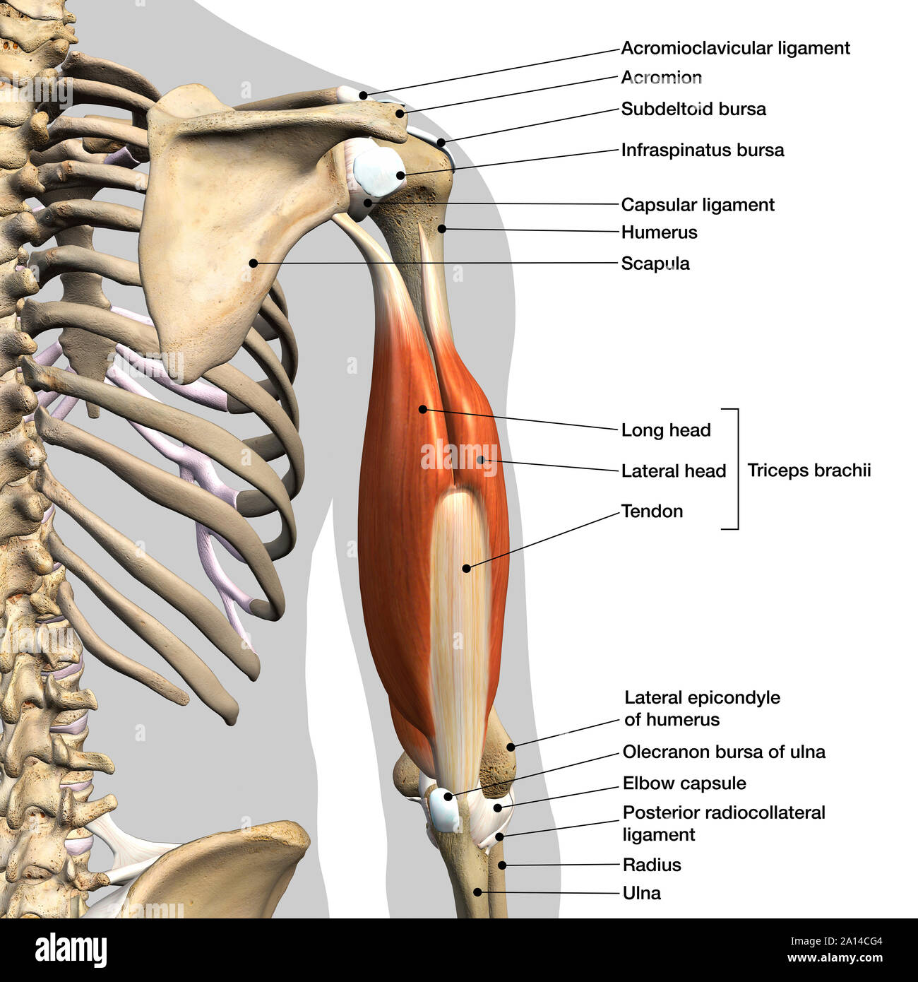
Labeled Anatomy Chart Of Male Triceps Muscles Connective Tissue And Bones On White Background Stock Photo Alamy from c8.alamy.com The shoulder muscles produce the characteristic shape of the shoulder and can be classified into two groups: Upper limb activities require a repetitive movement of the shoulder external rotator and abductor muscles. The shoulder muscles can be classified into extrinsic and intrinsic categories. They produce the characteristic shape of the shoulder, and can be divided into two groups Muscles of the rotator cuff labeled on a sagittal mr slice. Test your knowledge in our quiz about the shoulder muscles. An example of shoulder flexion can be seen when reaching forward to grasp an object. Specifically, the four rotator cuff muscles.
The muscles in the shoulder aid in a wide range of movement and help protect and maintain the main shoulder joint, known as the glenohumeral joint.
Due to its location, it belongs to the chest. The human shoulder is made up of three bones: Click on image for larger labeled, picture. An mri of the shoulder of a healthy subject was performed in the 3 planes of space (coronal, axial, sagittal) commonly used in osteoarticular imaging, with two weightings to explore the musculoskeletal. Diagram of human shoulder muscles / shoulder anatomy campbell hand shoulder surgeon southampton : Body muscles parts name 12 photos of the body muscles parts name body muscle. In human anatomy the shoulder joint comprises the part of the body where the humerus attaches to the scapula and the head. Upper limb activities require a repetitive movement of the shoulder external rotator and abductor muscles. Each muscle of the shoulder assists with specific movements. Ankle muscles diagram, back muscles diagram, chest muscles diagram, diagram of shoulder muscles and tendons, hip muscles diagram, knee muscles diagram, neck muscles diagram, rotator cuff muscles diagram, human. Tutorials on the shoulder muscles (e.g rotator cuff muscles: Want to learn more about it? One of the most important of these for.
That action is accomplished primarily by the combined actions of the deltoid muscle in the uppermost extent of the arm, the pectoralis major. They produce the characteristic shape of the shoulder, and can be divided into two groups One of the most important of these for. Test your knowledge in our quiz about the shoulder muscles. Shoulder muscles diagram includes some organs and can give you some detailed information as well as can be seen through shoulder anatomy diagram.
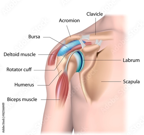
Physical Solutions Signs You May Have A Labrum Tear In Your Shoulder Physical Solutions from as1.ftcdn.net Related online courses on physioplus. The muscles in the shoulder aid in a wide range of movement and help protect and maintain the main shoulder joint, known as the glenohumeral joint. Upper limb activities require a repetitive movement of the shoulder external rotator and abductor muscles. Many of the muscles that attach onto the shoulder girdle also attach onto the: Muscles connecting upper limb to vertebral column muscles of shoulder. Diagram of human shoulder muscles / shoulder anatomy campbell hand shoulder surgeon southampton : Human muscles enable movement it is important to understand what they do in order to diagnose sports injuries and prescribe rehabilitation exercises. The latissimus dorsi also transversely extends and flexes the.
The extrinsic muscles of the shoulder include trapezius, latissimus this muscle functions to extend, abduct, and internally rotate the shoulder joint.
Test your knowledge in our quiz about the shoulder muscles. The shoulder is not a single joint, but a complex arrangement of bones, ligaments, muscles, and tendons that is better bones of the shoulder girdle. As a group, they are responsible for stabilizing the shoulder joint. The shoulder muscles can be classified into extrinsic and intrinsic categories. 7 draw labeled diagram to show the anastomosis around scapula. Shoulder flexion is movement of the shoulder in a forward motion. Upper limb activities require a repetitive movement of the shoulder external rotator and abductor muscles. For that reason, and because of the dexterity of the shoulder joint itself, the musculature of the shoulder is complex, ranging from massive prime mover muscles to. Diagram of human shoulder muscles / shoulder anatomy campbell hand shoulder surgeon southampton : They produce the characteristic shape of the shoulder, and can be divided into two groups Supraspinatus, infraspinatus, ters minor,.et), using interactive animations and labeled diagrams. Many of the muscles that attach onto the shoulder girdle also attach onto the: Because the thin actin filaments have overlapped there is a reduced.


0 Comments