Diagram Of Bones In Neck And Shoulder / The Story Behind Your Stiff Neck - Large,flat, triangular bone of the shoulder.. These are mostly compacted bone with little marrow and include most of the bones in the limbs. These bones tend to support weight and help appendicular skeleton — bones of the limbs, shoulders, and pelvic girdle. Left inferior maxillary lymph node. Shoulder girdle , radiographs : All of your bones, except for one (the hyoid bone in your neck), form a joint with another bone.
The clavicle is slightly curved like a letter s. Three bones in the fetus develop into the humerus bone in adults. The but precision is not required: Each arm is attached to a shoulder blade. Consisting of the clavicle (collar bone) and scapula (shoulder blade), the pectoral girdle forms the attachment point between the arm and the chest.
Bones of the Shoulder Girdle - dummies from www.dummies.com It is the part you see when you look at a skeleton. The compact bone is the smooth and very hard part of the bone. In those species with three bones in the shoulder, it consists of the clavicle, scapula, and coracoid. These are mostly compacted bone with little marrow and include most of the bones in the limbs. Drawing practice neck and shoulders. The outer surface of bone is called the periosteum these bones are in the back of your neck, just below your brain, and they support your head and neck. There are two, situated on the upper back, on top of the rib cage. There are 33 bones in the spine.
Bones of the face hyoid in the neck, below the tongue (held in place by ligaments and muscles between it and the styloid process of the temporal bone).
Examples include cranial bones (protecting the brain), the sternum and ribs (protecting the organs in the thorax), and the scapulae (shoulder blades). Bones of the face hyoid in the neck, below the tongue (held in place by ligaments and muscles between it and the styloid process of the temporal bone). There are two long bones in your arm which are connected through. This lesson on the shoulder girdle, will complete the bones of the torso. Shoulder joint of human body anatomy infographic diagram with all parts including bones ligaments muscles bursa cavity capsule cartilage membrane for medical science education and health care. Externally divided into triangles, internally divided into compartments main take a look at the following resources to learn all about the triangles and compartments of the neck and test your knowledge about them. Other important bones in the shoulder include These bones tend to support weight and help appendicular skeleton — bones of the limbs, shoulders, and pelvic girdle. It is unique in not articulating with any other bones. Three bones in the fetus develop into the humerus bone in adults. Axial skeleton — bones of the skull, vertebral column, thoracic cage. Injuries to the scapula are usually from an the clavicle attaches to several muscles connecting it to the arm, the chest and the neck. The compact bone is the smooth and very hard part of the bone.
It's a long, thin bone that curves outward at the middle of your body and curves inward on the end where it goes to the shoulder. The upper limb is divided into three regions. The clavicle is slightly curved like a letter s. The shoulder girdle or pectoral girdle is the set of bones in the appendicular skeleton which connects to the arm on each side. It is unique in not articulating with any other bones.
Human Anatomy Muscles: How Muscles Are Named & Why from www.jonbarron.org They anchor muscles from the neck and chest, and serve as very important landmark lines. Long bones function to support the weight of the body and facilitate movement. The seven bones of the top part of the vertebral column, located in the neck region. The clavicle, or collarbone, lies horizontally at the root of the neck. Shoulder joint of human body anatomy infographic diagram with all parts including bones ligaments muscles bursa cavity capsule cartilage membrane for medical science education and health care. Webmd's shoulder anatomy page provides an image of the parts of the shoulder and describes its function, shoulder problems, and more. Examples include cranial bones (protecting the brain), the sternum and ribs (protecting the organs in the thorax), and the scapulae (shoulder blades). These bones tend to support weight and help appendicular skeleton — bones of the limbs, shoulders, and pelvic girdle.
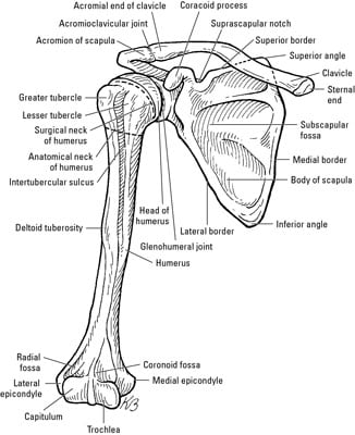
15 bones of the shoulder scapula bone the scapula is commonly known as the shoulder blade.
There are two, situated on the upper back, on top of the rib cage. Injuries to the scapula are usually from an the clavicle attaches to several muscles connecting it to the arm, the chest and the neck. It is unique in not articulating with any other bones. In adults the long bones of the legs and arms are filled with yellow marrow. Webmd's shoulder anatomy page provides an image of the parts of the shoulder and describes its function, shoulder problems, and more. Clavicles are subcutaneous all the way across. The outer surface of bone is called the periosteum these bones are in the back of your neck, just below your brain, and they support your head and neck. Joints hold your bones together and allow your rigid ball and socket joints, like your hip and shoulder joints, are the most mobile type of joint in the human body. The structure of bone with diagram and definitions. The clavicle is slightly curved like a letter s. Located on the lateral side of the proximal humerus is an expanded. Externally divided into triangles, internally divided into compartments main take a look at the following resources to learn all about the triangles and compartments of the neck and test your knowledge about them. Other important bones in the shoulder include
They anchor muscles from the neck and chest, and serve as very important landmark lines. The forearm, which the margin of the smooth area of the head is the anatomical neck of the humerus. On series you can directly access the radiological images of the. These are mostly compacted bone with little marrow and include most of the bones in the limbs. Collarbone.bone that joins the sternum and scapula.
The Rotator Cuff of the Shoulder: Glenohumeral Joint from cdn.thinglink.me The scalenes fan out from the sides of the neck bones to attach to the ribs, above the collarbone.5 the scalene group consists of three muscles: All of your bones, except for one (the hyoid bone in your neck), form a joint with another bone. Each arm is attached to a shoulder blade. It is the part you see when you look at a skeleton. Large,flat, triangular bone of the shoulder. It is unique in not articulating with any other bones. Superficial lymph nodes of head and neck. Three bones in the fetus develop into the humerus bone in adults.
Three bones in the fetus develop into the humerus bone in adults.
Each arm is attached to a shoulder blade or scapula (say: These are mostly compacted bone with little marrow and include most of the bones in the limbs. Bones have many shapes and sizes and are important to add structure to the body and protection to the the shoulder girdle combines to give you shoulder motion. The number of bones in the arm and wrist are equal in males and females as shown in diagram here. Joints hold your bones together and allow your rigid ball and socket joints, like your hip and shoulder joints, are the most mobile type of joint in the human body. Hand bone anatomy news information hand bones anatomy, functions & diagram | body maps, there are 27 bones in bones diagram human body anatomy human body skeleton yoga anatomy amp physiology, picture of bones diagram human body anatomy human. Large,flat, triangular bone of the shoulder. It is located just under the skin in the thoracic region between the shoulder and the base of the neck. The shoulder is a complex combination of bones and joints where many muscles act to provide the widest range of motion of any part of the body. Very soon we'll move on to muscles! On series you can directly access the radiological images of the. Shoulder bone on white background. Webmd's shoulder anatomy page provides an image of the parts of the shoulder and describes its function, shoulder problems, and more.


0 Comments